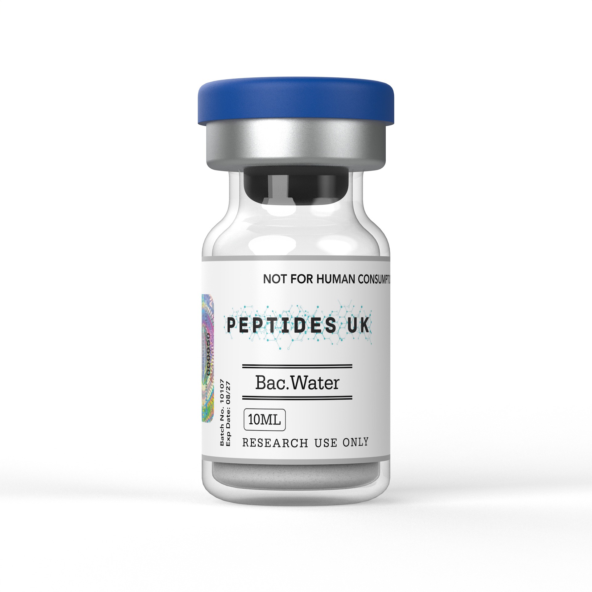
How to reconstitute peptides is a routine question in laboratory practice, because most research peptides are supplied as freeze-dried powders that must be dissolved before use in any assay. Reconstitution means returning a lyophilised peptide to solution at a defined concentration, using a solvent chosen for the sequence’s charge and hydrophobicity. Done poorly, it causes incomplete dissolution, aggregation, oxidation or inaccurate concentrations, any of which can compromise an experiment. This article explains the chemistry of dissolution, the published literature on peptide stability, the laboratory contexts in which stock solutions are prepared, and the purity, storage and handling standards expected in UK research settings. It concerns in-vitro work only.
How to reconstitute peptides: what the term means
In a laboratory context, reconstitution is the preparation of a stock solution from a lyophilised peptide. Lyophilisation, or freeze-drying, removes water by sublimation under vacuum after the peptide solution has been frozen. The technique was developed in the 1930s for preserving biological materials such as serum, and it remains the standard way to ship peptides because the dry state suppresses hydrolysis, oxidation and microbial growth. The result is usually a white, fluffy cake or a thin film at the base of the vial. The cake can be very small, and some of it may be dispersed on the vial walls or the stopper during transport.
The powder is not pure peptide. Solid-phase peptide synthesis, introduced by Bruce Merrifield in 1963, ends with cleavage from the resin using trifluoroacetic acid, so most research peptides are supplied as trifluoroacetate salts. Residual water and counter-ions mean that the net peptide content is often between 60 and 90 per cent of the gross weight. A researcher who weighs 1 mg of powder therefore does not hold 1 mg of peptide. This matters for every concentration calculation, and it is the first reason why the certificate of analysis should be read before the vial is opened.
Peptides are classified by sequence, and it is the sequence that determines how reconstitution should proceed. A short, charged, hydrophilic peptide may dissolve instantly in sterile water. A long, hydrophobic sequence with few charged residues may need an organic co-solvent. Peptides containing cysteine, methionine or tryptophan need additional care to limit oxidation. Because no single protocol suits every sequence, the question of how to reconstitute peptides is best answered by a systematic approach to solubility rather than a fixed recipe.
Mechanism of action: the chemistry of dissolution
Dissolution is governed by the interaction between the peptide’s side chains, its terminal groups and the solvent. Water is a good solvent for peptides that carry enough charged or polar residues to form favourable hydrogen bonds and ion-dipole interactions. Hydrophobic residues such as leucine, isoleucine, valine, phenylalanine and tryptophan do the opposite. They favour peptide-peptide contacts over peptide-water contacts, which promotes aggregation and slows or prevents dissolution.
Net charge at a given pH is the most useful predictor of solubility. A practical estimate assigns +1 to each lysine, arginine and the free N-terminus, and −1 to each aspartic acid, glutamic acid and the free C-terminus. Histidine is approximately neutral at physiological pH but becomes positively charged below about pH 6. A peptide with a positive net charge is described as basic and generally dissolves best in water or a dilute acid such as 10 per cent acetic acid. A peptide with a negative net charge is acidic and generally dissolves best in water or a mildly basic solution such as dilute ammonium bicarbonate. A peptide with a net charge near zero is the most difficult, because it is least soluble near its isoelectric point, and it often requires a small volume of organic solvent such as dimethyl sulfoxide or dimethylformamide before it is diluted into the aqueous buffer.
Co-solvent choice has chemical consequences. Dimethyl sulfoxide can oxidise cysteine and methionine side chains, so dimethylformamide or acetonitrile is often preferred for those sequences. Dilution should be dropwise into the aqueous phase with gentle mixing, because rapid addition can cause local precipitation. Sonication for short bursts in a cool water bath can help break up particles, although heat and prolonged energy input can damage sensitive sequences.
Once dissolved, the peptide faces competing degradation pathways. Deamidation of asparagine and glutamine, aspartate isomerisation, oxidation, hydrolysis at aspartyl bonds and physical aggregation all proceed faster in solution than in the dry state. Their rates depend on pH, temperature, buffer species and the neighbouring residues. Understanding these mechanisms explains why most protocols specify a slightly acidic sterile buffer, typically between pH 5 and 6, why concentrated stocks are aliquoted and frozen, and why working solutions are prepared fresh where possible.
What the research shows
The published literature relevant to reconstitution is largely physical-chemical, not pharmacological. The following four research areas are described in general terms, and readers should consult the primary papers directly.
Merrifield’s 1963 paper in the Journal of the American Chemical Society described solid-phase synthesis of a model tetrapeptide on a polymer support, with stepwise coupling and washing. Its relevance here is practical. The cleavage and purification chemistry that followed explains why research peptides carry counter-ions and residual moisture, and why net peptide content must be considered when preparing a solution. The method’s development can be traced through a PubMed search on solid-phase peptide synthesis.
A substantial review literature covers the science of freeze-drying proteins and peptides. Wang’s 2000 review in the International Journal of Pharmaceutics is a frequently cited example. These reviews describe how freezing and drying stresses affect biomolecules, how residual moisture influences storage stability, and why excipients such as sugars are used in some formulations. They support the general principle that lyophilised material is far more stable than the same peptide in solution. Related papers can be found through a PubMed search on lyophilisation and peptide stability.
Studies of chemical degradation in solution, including work by Geiger and Clarke published in the Journal of Biological Chemistry in 1987, used synthetic model peptides in buffered solutions to examine deamidation and isomerisation at asparagine and aspartate residues. These studies reported that the reactions proceed through a cyclic succinimide intermediate, and that rates depend on pH, temperature and the identity of the neighbouring residue. The practical implication is that sequences containing asparagine followed by glycine, for example, are particularly prone to degradation once dissolved.
Research on aggregation, including reviews such as Bondos and Bicknell’s 2003 paper in Analytical Biochemistry, addresses how concentration, pH, ionic strength and handling influence the formation of aggregates before, during and after purification. The findings support cautious practice during reconstitution, including gentle mixing, avoidance of vigorous vortexing that introduces air-liquid interfaces, and avoidance of repeated freeze-thaw cycles.
None of these studies concerns any effect in humans, and none should be read as such.
Research applications
Stock solutions prepared by reconstitution are the starting point for a wide range of laboratory work. In receptor pharmacology, peptide ligands are dissolved and serially diluted to construct concentration-response curves in transfected cell lines, where accurate stock concentration is essential for calculating affinity constants. In enzymology, peptide substrates and inhibitors are prepared as concentrated stocks and diluted into assay buffers for kinetic measurements, including fluorogenic protease assays in which small errors in substrate concentration distort the calculated kinetic parameters.
In immunology, peptide libraries covering a protein’s full sequence are reconstituted, often in a small volume of dimethyl sulfoxide followed by dilution into culture medium, and used to stimulate T-cell responses in vitro. Here the final co-solvent concentration is controlled carefully, because organic solvents can affect cell viability. In biophysics and structural biology, reconstituted peptides are used in circular dichroism and nuclear magnetic resonance experiments to study secondary structure and folding, where aggregation or buffer incompatibility can produce misleading spectra.
In analytical work, reconstituted peptides serve as reference standards for liquid chromatography and mass spectrometry. These applications depend heavily on accurate concentration, which in turn depends on correct handling of net peptide content. Across all of them, a consistent, documented reconstitution procedure is part of good experimental practice, because it removes one source of variability between replicates and between laboratories.
Purity, storage and handling
Purity is verified principally by reversed-phase high-performance liquid chromatography with ultraviolet detection at 214 nm, where the peptide bond absorbs. The chromatogram shows the main peak and any impurities such as deletion sequences, truncated chains and incompletely deprotected products. Mass spectrometry confirms that the observed molecular mass matches the theoretical value. Together they establish identity and purity, and a purity figure supplied without chromatographic data is difficult to verify independently.
A batch-specific certificate of analysis matters because synthesis is not perfectly reproducible. Impurity profiles, counter-ion content and water content differ between production runs, and these differences feed directly into the concentration of any stock solution. A generic specification sheet cannot capture them, whereas a document tied to a lot number can.
Lyophilised peptides are best stored at −20 °C, with −80 °C preferred for long-term archives or sensitive sequences. Before opening, the vial should be allowed to reach room temperature in a desiccator so that condensation does not introduce moisture to the powder, and a brief centrifugation will bring any dispersed material to the base. Light can accelerate oxidative damage, particularly in sequences containing tryptophan, tyrosine or methionine, so amber vials or storage in the dark are sensible precautions.
Because degradation accelerates once a peptide is in solution, batch-level HPLC verification matters more at the point of reconstitution than it does for stable reagents, and UK laboratories assessing how to buy best peptides should expect a batch-specific certificate of analysis and lyophilised storage at −20 °C. Reconstituted stock solutions are best prepared at a defined molar concentration, commonly in the low millimolar range or around 1 to 2 mg/mL for assay work, using the net peptide content from the certificate of analysis to calculate the amount of solvent required.
The procedure itself is simple but benefits from discipline. A small test portion should be dissolved first where material is limited and the sequence is unknown, so that an unsuitable solvent does not consume the entire batch. The solvent should be added slowly down the side of the vial and the contents mixed by gentle swirling or slow inversion, not by vigorous shaking. If the solution remains cloudy, a brief period of sonication or the stepwise addition of a stronger solvent may be tried, and a solution that remains turbid should be centrifuged before use. For biological assays, a 0.22 µm low-protein-binding filter can remove particulates, although some peptide may be lost to the membrane, so filtration is usually performed at higher concentration and the loss accounted for.
Stock solutions should be divided into single-use aliquots in low-binding tubes, because peptides readily adsorb to glass and some plastics, and then frozen promptly. Repeated freeze-thaw cycles promote aggregation and degradation, so each aliquot should be thawed once and discarded after use. Short-term storage at 2 to 8 °C is acceptable only for sequences known to be stable in solution, and the period should be kept brief. Solvent and pH should be chosen with the sequence in mind, favouring slightly acidic conditions for most peptides and avoiding basic pH for those containing cysteine, which oxidises more quickly as pH rises.
Standard laboratory practice applies throughout. Gloves and eye protection should be worn, and each vial and aliquot should be labelled with the sequence, lot number, concentration, solvent and date of reconstitution. Waste, including organic co-solvents, should be disposed of in line with institutional chemical-waste procedures.
Frequently asked questions
How do you reconstitute peptides for laboratory assays?
Allow the vial to reach room temperature, centrifuge briefly, then add a solvent suited to the sequence’s net charge, usually sterile water, dilute acetic acid or a small volume of organic co-solvent. Mix gently, calculate concentration from net peptide content, aliquot into low-binding tubes and freeze.
What solvent should be used to dissolve a peptide?
Sterile water suits many short, charged sequences. Basic peptides often dissolve in dilute acetic acid, and acidic peptides in dilute ammonium bicarbonate. Hydrophobic sequences may need dimethylformamide or dimethyl sulfoxide first, then dilution. Cysteine- or methionine-containing peptides generally avoid dimethyl sulfoxide because of oxidation risk. Always test a small portion first.
How long are reconstituted peptides stable?
Stability depends on sequence, solvent, pH and temperature, so no single figure applies. Solutions degrade far faster than lyophilised powder. Frozen single-use aliquots generally last longest, whereas refrigerated solutions are suitable only for short periods. Sequences with asparagine, methionine or free cysteine need particular caution and should be checked against sequence-specific literature.
Why will my peptide not dissolve?
Poor solubility usually reflects hydrophobic content, a net charge near zero at the chosen pH, or aggregation. Adjusting pH away from the isoelectric point, adding a small amount of organic co-solvent, or using brief sonication often helps. Persistent insolubility may indicate a problem with the batch, so compare against the certificate of analysis.
Peptides are supplied for in-vitro laboratory research only. They are not for human or veterinary use, and nothing in this article should be read as guidance on administration, dosing or any effect in a living organism.